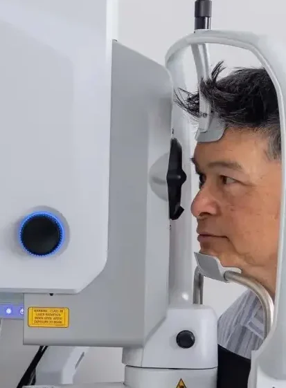
Precise imaging to support accurate diagnosis and care.

Advanced Retina Diagnostics

Expert care. Clear answers. A patient-centered approach to retina health.

img 9903
Services

Specialized care for your retina and vision.

From precise diagnostic imaging to medical and surgical treatment, every aspect of your care is guided by clinical expertise, thoughtful evaluation, and a clear understanding of your individual needs.

Eye Examinations · Retinal Diagnostics · Laser & Injections · Retina Surgery

A complete look at your eye health and vision.

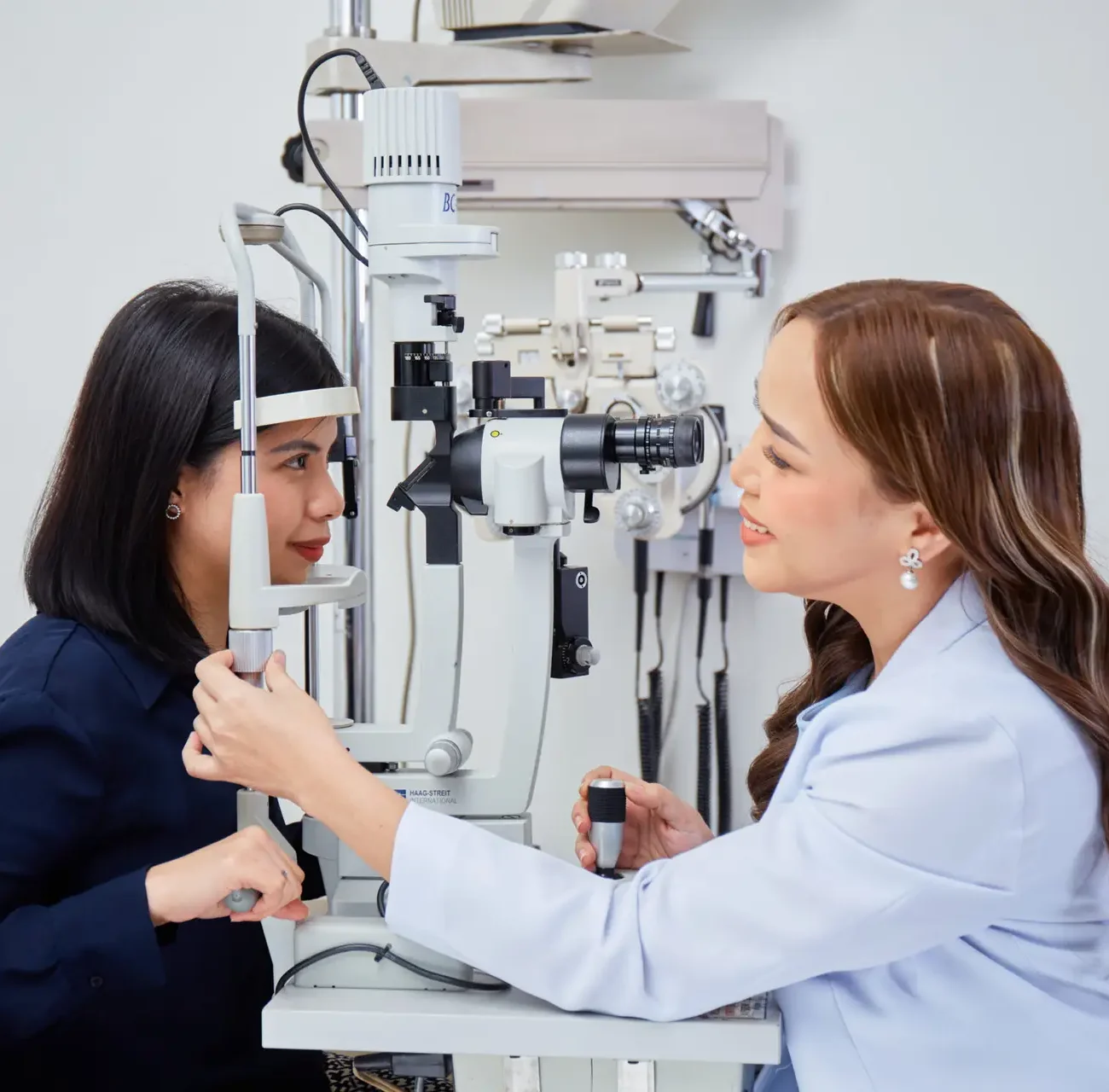
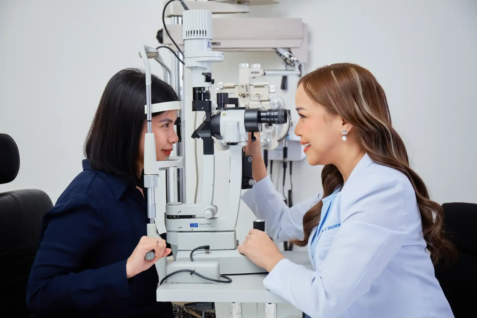
Comprehensive Eye Examination

A comprehensive eye exam goes beyond checking your glasses prescription. Dr. Patty carefully assesses the overall health of your eyes, looking for signs of eye conditions that may not yet affect your vision. With a clearer understanding of your eye health, you can make informed decisions about your care and take the right steps to protect your sight.
img 9894
oct scan (1)
A detailed scan of the retina.

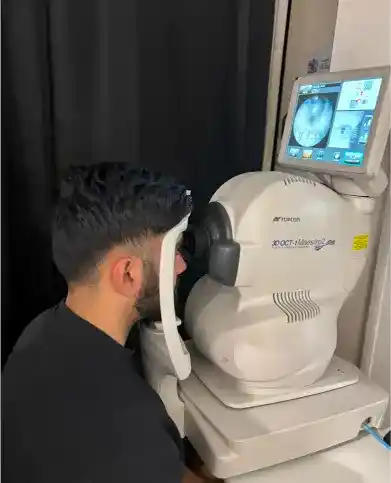
Optical Coherence Tomography (OCT)


OCT creates detailed, cross-sectional images of the retina, allowing your doctor to see its individual layers. It is commonly used to detect and monitor conditions such as macular edema, macular holes, AMD, retinal detachments, and other retinal conditions.

A detailed look at blood flow in the retina.

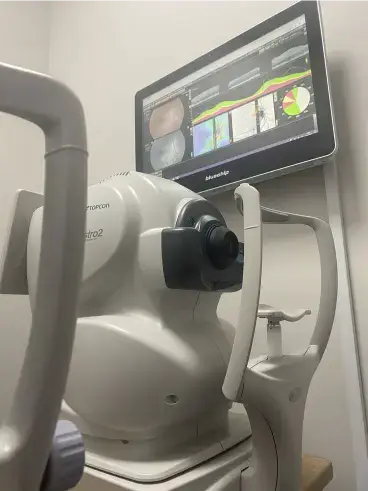
OCT Angiography (OCT-A)


OCT creates detailed, cross-sectional images of the retina, allowing your doctor to see its individual layers. It is commonly used to detect and monitor conditions such as macular edema, macular holes, AMD, retinal detachments, and other retinal conditions.

oct scan (1)
0dd3efa414b175d9ab6198a2daf3a308
A detailed photograph of the retina.

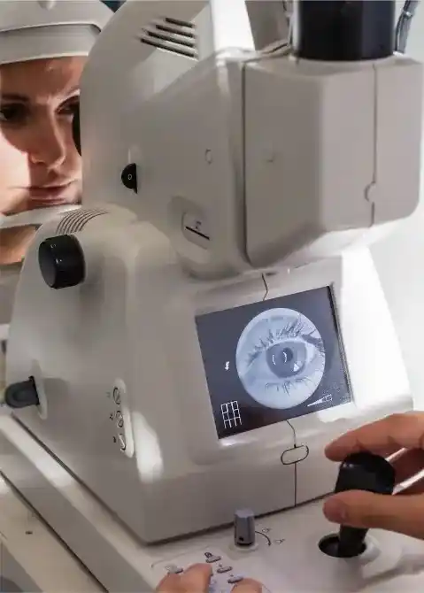
Fundus Photography / Wide-Field Retinal Imaging

Specialized cameras capture images of the back of the eye. Wide-field imaging can show a much larger area of the retina, including the peripheral retina, helping your doctor document and monitor retinal changes over time

A closer look at retinal blood vessels.

Fluorescein Angiography (FA)

A fluorescent dye is injected into a vein, usually in the arm, while a special camera captures images as the dye travels through the retinal blood vessels. This can help identify areas of leakage, blockage, or abnormal blood vessel growth.

3 fundus (1)
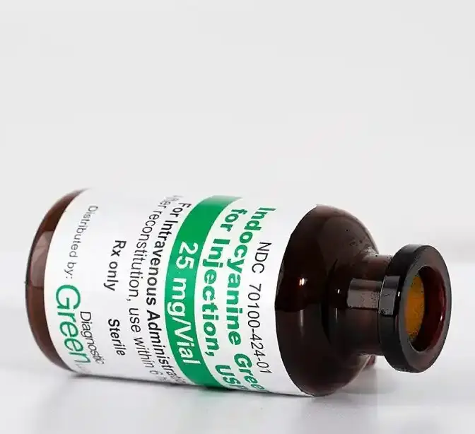
icg dye and sterile water with open bottle 1536x614 (2)
A deeper look at the retinal and choroidal circulation.

Indocyanine Green Angiography (ICGA)

ICGA uses an injected dye to provide detailed images of the blood vessels beneath the retina, particularly within the choroid. It can be especially useful when evaluating certain retinal and choroidal conditions.

An image that reveals changes in the retinal pigment layer.

Fundus Autofluorescence (FAF)

A fluorescent dye is injected into a vein, while a camera captures images as it travels through the retinal blood vessels. FAF is a noninvasive test that maps the retinal pigment epithelium, aiding in the diagnosis of retinal diseases.

65b6fcb5e4c1ae76f30cb256 6576976a883c9d3bd2f81c86 zeiss clarus retinal photography fundus autofluorescence faf macular dystrophy adelaide